Sperm are so small that they are impossible to see with the naked eye. They are tiny wrigglers that carry 23 chromosomes to fertilize an egg.
If all the sperm in one ejaculate lined up end to end, they’d stretch six miles – This section is authored by the website’s editor XXX Teens Sex. Scientists like Pitnick have found a way to see these sperm cells.
Head
A sperm is a mature, haploid gamete that carries genetic information. It is a single cell with three distinct parts: a head piece, a midpiece, and a tail that propels it for swimming and helps breach an egg (Dcunha, 2022). It is produced in small tubes within a man’s testicles called the seminiferous tubules. (Dcunha, 2022)
The head of the sperm contains its nucleus full of paternal genetic material and cytoplasm (a salt-water-protein fluid that fills cells). It also has a specialized structure on the tip called an acrosome which is bound to a protein that allows the sperm to digest and break open the egg for fertilization.
The acrosome is also present on the midpiece of the sperm. The midpiece is rich in mitochondria that produce ATP, an energy molecule. It is the sperm’s power source. At the very end of the sperm is the tail, which has a whip-like shape that helps it swim and then to breach an egg. The entire sperm is about 0.05 millimeters long and contains a variety of nutrients like vitamin B12, creatine, cholesterol, citric acid, and various salts and enzymes (Dcunha, 2022). A smartphone microscope would be useful in countries where normal laboratory equipment is too expensive or unavailable, says Stuart Lavery, a urologist at Hammersmith Hospital in London.
Neck
Sperm cells, or spermatozoa, have an expanded head, a narrow neck and a long principal tail. They are located in the testicular organs of male human beings and animals, where they are formed from spermatocytes through a process known as spermiogenesis. These sperm cells are then transported to the penis through the seminiferous tubules where they develop into mature sperm.
Inside the sperm head is a nucleus, which contains genetic information, and cytoplasm, a fluid filled with salt, water and protein. A specialized structure at the tip of the head, called the acrosome, contains hydrolytic enzymes that may help the sperm penetrate the outer shell of the female egg and fertilize it.
The sperm cell’s motile flagellum consists of a central axoneme surrounded by nine evenly spaced doublet microtubules. It is this flagellum that propels the sperm to swim and breach the woman’s egg to fertilize it.
Observing sperm cells under a microscope is a common part of routine semen analysis done to detect issues with the fertility of men. The sperm cells are evaluated for their normal morphology, which includes head size and appearance, midpiece structure and the length of the tail. Sperm that have a normal morphology are able to swim in a straight line and have healthy DNA that can fertilize the egg. Sperm with abnormal morphology, such as macrocephaly (giant head), may have problems entering the woman’s egg and fertilizing it.
Middle
As we learned from biology class, sperm cells (also known as spermatozoa) contain chromosomes. When they combine with an egg cell, sperm and eggs join to form a diploid organism, resulting in the birth of a baby. These chromosomes are made up of DNA, which carries genetic information. The baby inherits half the chromosomes from its father and half from its mother.
To examine sperm under the microscope, you’ll need to prepare a semen sample by using a cotton swab to collect a smear of the sample on a glass slide. A smear is a thin layer of sample that contains sperm and other cellular debris. This sample can then be placed under a biological microscope for sperm evaluation and motility testing.
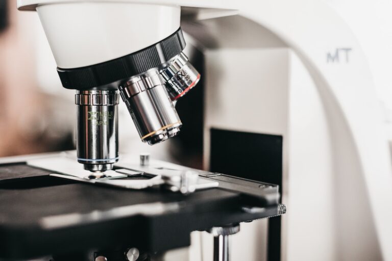
You can use either a phase contrast or Nomarski DIC microscope for sperm evaluation. Many professionals prefer a phase contrast microscope over staining because it’s less likely to damage or kill living cells, as is the case with staining.
A phase contrast microscope also makes it easy to see sperm without having to scan the slide manually, which is time consuming and can lead to mistakes. This type of microscope also offers the option for a digital camera attachment. This allows you to easily capture, save and monitor the movement of a sperm sample while under the microscope.
Tail
Sperm cells are far too tiny to see with the naked eye. They measure just 0.002 inch from head to tail, or 50 micrometers. And if you could get all the sperm in one ejaculate to line up end to end, they would stretch six miles.
That’s why a microscope is essential for assessing sperm motility, an important factor in livestock breeding. But a typical lab microscope with its normal objectives and lenses just won’t do the job. You need a specialized sperm microscope with the right magnification and other features to ensure that you can see sperm cells in detail and assess their movement.
For example, a specialized sperm microscope with a digital camera will let you capture images and video footage of your samples while they’re still alive. You can then easily share these images with colleagues and specialists for consultations or further evaluation.
In terms of morphology, most sperm characterization is done using stained smears and light microscopy at 1000x magnification. But this method is limited and has certain drawbacks. For example, it is time consuming and requires a lot of manual scanning. The latest technologies, however, can help to speed up this process. They involve the automated scanning of microscope slides and then optimized image analyzing software. These systems can detect sperm cells, even those that are not stained with Sperm HYLITER.
See Also:


